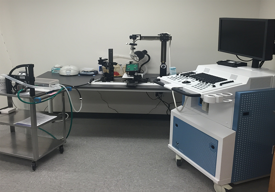
 Vevo-2100 Ultrasound Imaging System (VisualSonics): This advanced system includes a wide range of transducers with frequencies 15-50 MHz, which are ideal for “micro” ultrasound imaging of organs at depths up to 3 cm and frame rates up to 10,000 frames per second. Uses include:
- Cardiac function indices: As adult mice have heart rates of approximately 400 to 650 beats per minute without anesthesia, ECG-gated kilohertz visualization is used, which yields >1000 frames per second, for measuring 1) endocardial and epicardial areas at end-systole and end-diastole, and 2) left ventricle base to apex distance at end-systole and end-diastole. Based on these measurements, various indices of heart functions can be calculated, including fractional shortening (FS), ejection fraction (EF), stroke volume (SV), average wall thickness at end-diastole, and LV mass.
- Vessel diameter and flow profile: The transmitral flow profile is recorded in an apical 4-chamber view sound window using pulse-wave Doppler echocardiography to measure: 1) E- and A-wave peak velocities, 2) E-wave deceleration and acceleration time, 3) isovolumetric relaxation and contraction time, 4) ejection time, and 5) velocity time integral. Using these parameters, the E/A ratio and the myocardial and valvular performance index are. The diameters of the ascending, transverse and lower abdominal aorta, and the innominate and carotid arteries are measured at end-diastole and at end-systole. Velocity, pattern, and direction of flow in arteries, veins, and possibly lymphatics are measured using the pulse-wave, and color-Doppler flow modes.
- A novel method of blood collection: The Core has developed a reliable method of plasma preparation with minimal platelet activation by obtaining blood from the left ventricle inflow tract of anesthetized mice under ultrasound guidance. This technique consistently yields virtually no platelet activation, as demonstrated by the very low levels of platelet α-granule proteins TGFβ1, TSP1, and PF4 in plasma relative to the high levels in plasma from conventional retrobulbar blood drawing.
- Vascular perfusion, thrombus detection, organ/tumor volume, and prenatal embryo analysis: The Vevo-2100 provides rapid identification of vessels, flow rates, velocity and direction in a very sensitive and automated set up for thrombosis detection in coronary arteries of the heart, carotid and kidney, or in small vessels such as femoral arteries. The device also includes a 3D image mode that captures 3-dimensional images of virtual sections in all directions, x-, y-, z- and plane variations, and provides accurate volume measurement of tissues and tumor growth. It provides linear contrast imaging using ultrasound-based contrast agents, which can precisely measure vascular blood flow and provide detailed analysis of tissue and tumor perfusion using the Vevo-Vascular Analysis software, which is designed to measure vessel wall anatomy and motion in small-animal models (mice, rats, rabbits, zebrafish, and fly). It is also capable of ultrasound-guided microinjection in embryos and of precise prediction of early embryonic age and phenotype, which is critical for many investigators who focus on vessel structures and functions during embryonic development.
- Novel myocardial infarction model: The Core is developing a mouse model of myocardial infarction by introducing platelet microthrombi into the left ventricle. Preliminary data show that some of the microthrombi enter and lodge within the coronary arteries and cause myocardial infarction as measured by defective coronary artery blood flow and impaired heart function by the Doppler blood flow velocity and vessel diameter measurement for shear calculation.
- Transverse aortic constriction model: This model enables study of pressure overload-induced cardiac hypertrophy and heart failure. The model has been used to document platelet TGFb1-induced cardiac hypertrophy and fibrosis and can be used for many other indications.
Hemavet 950 FS: The analyzer automatically quantifies red blood cells, white blood cells, and platelets in the blood. In addition, it provides a detailed breakdown of the blood, including hemoglobin, neutrophils, lymphocytes, monocytes, eosinophils, basophils, platelets, and packed cell volume.
IDEXX VetLab® Chemistry Analyzer: Altered blood chemistry is often present in mouse models with cardiovascular dysfunctions. This Core instrument measures electrolytes, plasma proteins, lipid profiles, heart/liver enzymes, and kidney functions.
Small Animal Metabolic Screening System: Pathologic changes in metabolism are risk factors for many cardiovascular diseases. Conversely, cardiovascular disease conditions may induce metabolic disorders. Being able to characterize changes in animal metabolism that promote or result from cardiovascular pathologies provides an important resource for conducting mechanistic and translational cardiovascular research. The rodent metabolic system utilizes indirect calorimetry, body composition measurements, and equipment to induce and record locomotor activity to provide a comprehensive approach for phenotyping metabolism in mice and small rats.
DSI PhysioTel Telemetry System: This instrument measures cardiovascular functions in un-anesthetized animals during spontaneous activity and in response to stress challenges (e.g., exercise). The instrument includes PA-C10 Series Transmitters for in vivo measurement of pressures, including arterial and left ventricular, and an ETA-F10 Transmitter for in vivo measurement of ECG, temperature and activity data. The Core staff will work with investigators to implant, monitor, and analyze the biometric data and to optimize experimental designs.


